• Skip to primary navigation
  • Skip to main content
  • Departments
    • Aerospace Engineering
    • Bioengineering
    • Civil and Environmental Engineering
    • Electrical Engineering and Computer Sciences
    • Engineering Science
    • Industrial Engineering and Operations Research
    • Materials Science and Engineering
    • Mechanical Engineering
    • Nuclear Engineering
  • News
    • Berkeley Engineer magazine
    • Social media
    • News videos
    • News digest (email)
    • Press kit
  • Events
    • Events calendar
    • Homecoming
    • Cal Day
    • > Cal Day 2023 events
    • Commencement
    • View from the Top
    • Kuh Lecture Series
    • Minner Lecture
  • College directory
  • For staff & faculty
Berkeley Engineering

Educating leaders. Creating knowledge. Serving society.

  • About
    • Facts & figures
    • Rankings
    • Mission & values
    • Equity & inclusion
    • Voices of Berkeley Engineering
    • Leadership team
    • Milestones
    • Facilities
    • Maps
  • Admissions
    • Undergraduate admissions
    • Graduate admissions
    • Visit
    • Maps
    • Admissions events
    • K-12 outreach
  • Academics
    • Undergraduate programs
    • Majors & minors
    • Undergraduate Guide
    • Graduate programs
    • Graduate Guide
    • Innovation & entrepreneurship
    • Kresge Engineering Library
    • International programs
    • Executive education
  • Students
    • Advising & counseling
    • Programs
    • Academic support
    • Student life
    • Wellness & inclusion
    • Undergraduate Guide
    • Degree requirements
    • Forms & petitions
    • Resources
  • Research & faculty
    • Centers & institutes
    • Undergrad research
    • Faculty
  • Connect
    • Alumni
    • Industry
    • Give
    • Stay in touch
Home > News > Good for the image

Good for the image

Berkeley Engineer Spring 2020 magazine cover
April 14, 2020
This article appeared in Berkeley Engineer magazine, Spring 2020
Brain with hemorrhages highlightedscans

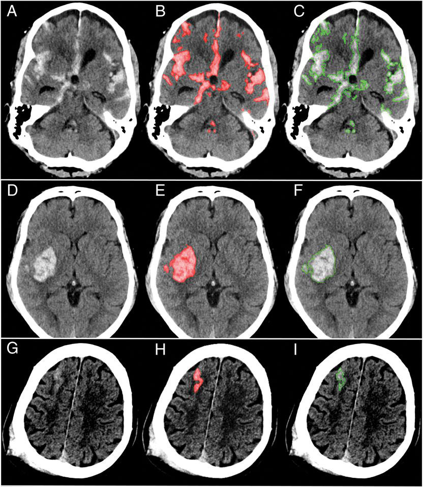
Images A, D and G are the original brain scans. The red shading on scans B, E and H indicates hemorrhages as recognized by the algorithm; the green outline on images C, F and I shows hemorrhages as determined by neuroradiologists. (Photos courtesy UCSF)

Radiologists can review thousands of brain images each day, searching for minute signs of traumatic brain injuries, strokes and aneurysms. Now, Berkeley researchers, working with scientists at UCSF, have developed a computer algorithm that could help radiologists focus on the most important images and examine them more closely. Using artificial intelligence to more efficiently and accurately recognize abnormal CT scans, the team’s algorithm took just one second to identify head scans with signs of hemorrhage — even some that experts missed. For each abnormality, the algorithm also provided a detailed outline, location and subtype, which could help physicians quickly determine the appropriate treatment. According to study co-author Jitendra Malik, Berkeley professor of electrical engineering and computer sciences, the researchers were able to use a small number of images to train the algorithm by using a type of deep learning known as a fully convolutional neural network, or FCN. In these training images, each small abnormality was manually delineated at the pixel level, helping to create an extremely accurate algorithm.

Topics: EECS, Health, Research, Robotics & AI
  • Contact
  • Give
  • Privacy
  • UC Berkeley
  • Accessibility
  • Nondiscrimination
  • instagram
  • twitter
  • linkedin
  • facebook
  • youtube
© 2023 UC Regents